Meet our CEO →
Healthcare Computer Vision Development Services
We build computer vision models that find, count and measure what matters in medical images, test them on data from sites they never saw, and run them where the data lives: in the browser, on a phone or inside a device.
Discuss your imaging project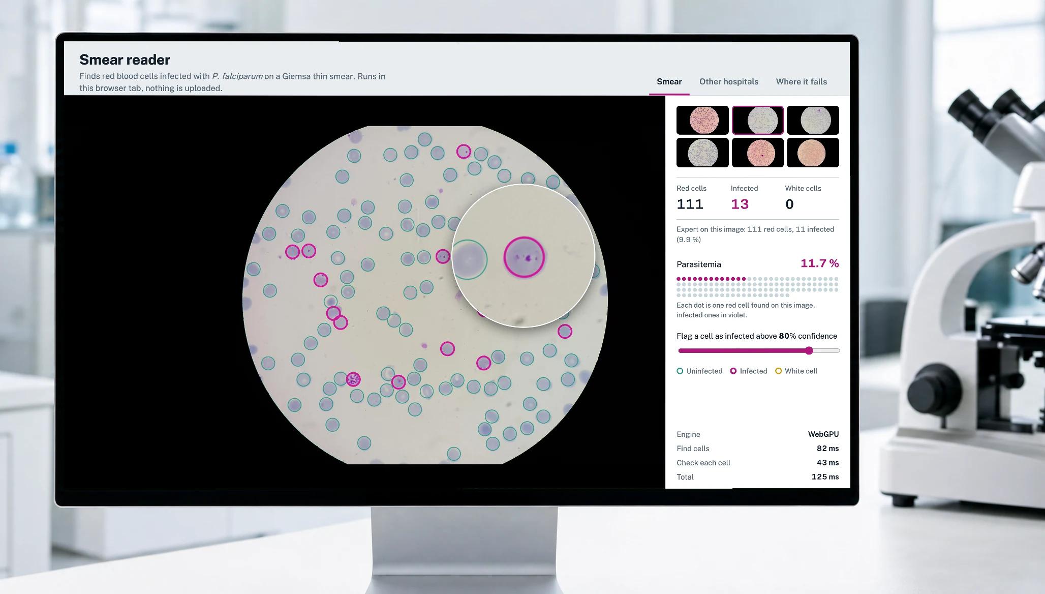
Computer vision that shows its work
Healthcare computer vision is software that reads medical images and video the way a trained eye does: it finds, counts and measures. Built into medical image analysis software, it turns slides, photos and scans into numbers a clinician or a lab can act on.
A model that scores well on its own test set can still fail in the next clinic: another camera, another stain, another labelling habit. We build medical computer vision the way a reviewer will check it, with patients held out from training, a test on data from another site, a threshold the clinical team controls, and a plain list of where the model fails.
Our own research: Smear reader
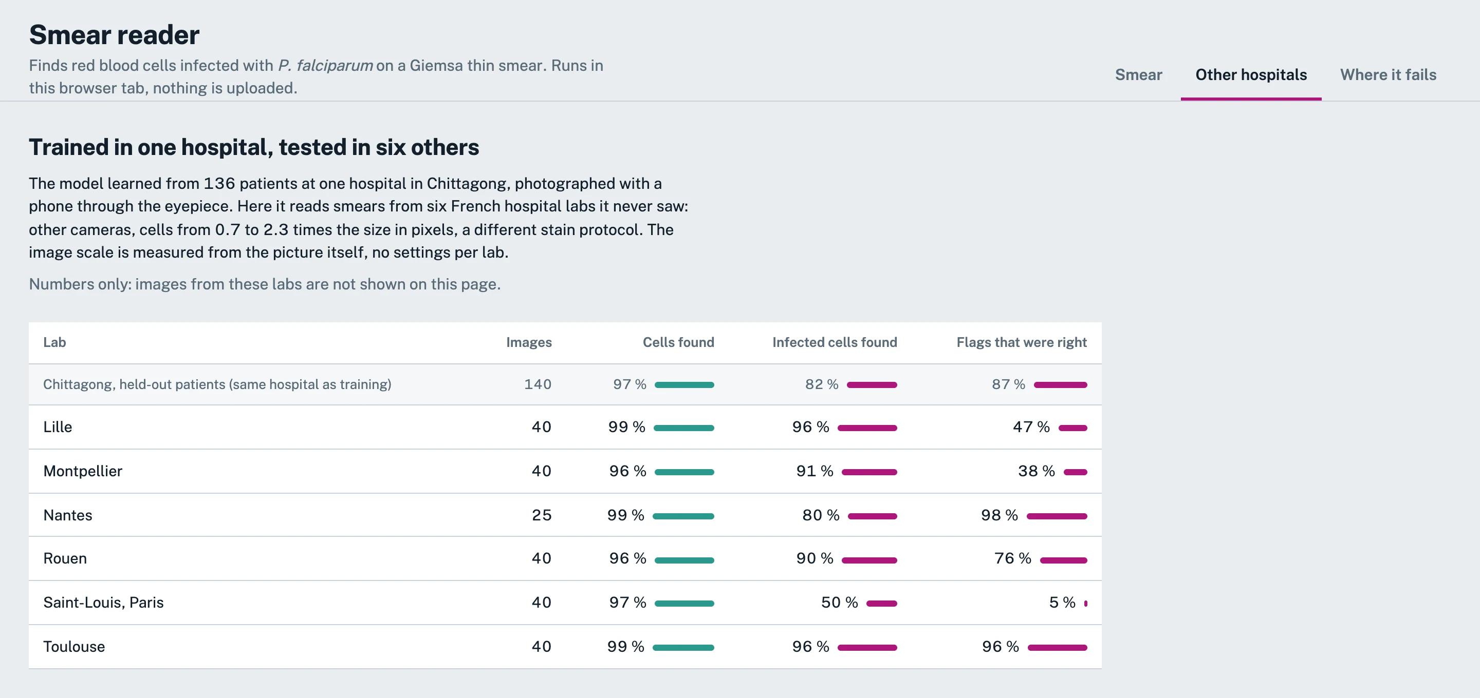
Smear reader is our own computer vision R&D: it finds malaria-infected red blood cells on a blood smear photo, right in the browser. We trained two small models from scratch on open data from one hospital (images courtesy of the U.S. National Library of Medicine), then tested them without retraining on six French hospital labs (Guemas et al., CC BY 4.0).
Read the case0.1 s
per 5312×2988 photo in the browser, with WebGPU
96–99%
of red cells found in six hospital labs the model never saw
0.98
correlation of parasitemia with the expert, per held-out patient
4.2 MB
for both models, running on the device
Our medical image analysis services
From a first look at your images to a model running inside your product.
Discuss your imaging project
Feasibility check on your images
We take a sample of your images and labels, build a baseline model and tell you what performance is realistic, which data is missing and what it would take to close the gap.

Custom model development
Detection, segmentation and classification models designed for your images, trained from scratch or from a pretrained backbone, and sized for the hardware they must run on.

Data and annotation strategy
Labelling rules, annotation tooling and quality checks, with data split by patient and by site, so the numbers you see today still hold next year.

Validation across sites
Tests on data from sites the model never saw, failure analysis image by image, and reports a clinical or regulatory reviewer can follow.

On-device and in-browser inference
Models exported to ONNX and run with WebGPU, WebAssembly or mobile runtimes, so images can stay on the device and results come back in a fraction of a second.

Integration into your product
The model inside your web or mobile app, lab system, PACS viewer or device software: reading DICOM, writing results back to the EHR, with the audit trail and monitoring around it.
EHR integration servicesComputer vision in healthcare: where it helps
Any task where a trained eye counts, measures or compares what it sees in an image is a candidate for computer vision.

Microscopy and digital pathology
Finding, counting and classifying cells and other structures on slides and smears, and measuring what a technician would otherwise count by hand.

Dermatology and wound photos
Measuring the area and change of a lesion or a wound from photos taken in the clinic or at home.

Radiology: X-ray, CT and MRI
Detecting and measuring findings on scans to prioritise a worklist and support the reading.

Ophthalmology
Grading retinal photos and flagging the ones that need a specialist’s attention first.

Point-of-care devices
Reading a result from a camera inside a device, so the device reports a number, not a picture.

Surgical and endoscopy video
Recognising instruments, phases and events in video, in real time or after the procedure.

Rehabilitation and remote monitoring
Tracking body pose and movement from a phone camera to score exercises at home.

Lab and pharmacy automation
Checking labels, counting items and confirming the right item is in the right place.
What we test for before a model meets a new clinic
Six checks that decide whether a model survives new data. Each one changed the numbers in our own research.
Discuss your imaging projectLabelling rules that differ between sites
In our malaria research, the French labs label cells cut by the frame edge and the training data does not. Counting that as a miss made a good detector look bad.

Stain, colour and lighting shift
Colour augmentation covers much of the gap between labs, but not all of it: the pinkest smears were the main source of missed infections.

Image scale and resolution
Cells came in 0.7 to 2.3 times the training size. The model measures the scale from each image, so no site needs its own settings.

Leakage between training and test
Several images of one patient look alike. We split by patient and by site, never by image, or the scores come out too good.

An imperfect reference
Some confident "false alarms" show a dot that looks like a parasite. One expert labelled the reference, so it caps the measured precision.

Rare positives and the threshold
When positives are rare, a default threshold floods the screen with flags. We tune it on separate patients and leave it in the user’s hands.
How we build a healthcare computer vision model
Technologies we work with
We pick the model and the runtime for where it has to run: a server, a phone, a browser tab or a device.
Deep learning

PyTorch

TensorFlow

Keras

MONAI

Hugging Face

Scikit-learn

Python
Images and video

OpenCV

Albumentations

Pillow

NumPy

SciPy

FFmpeg

GStreamer
Labelling and training

CVAT

Label Studio

Weights & Biases

MLflow

DVC
Deployment
ONNX
ONNX Runtime

Core ML

WebGPU

WebAssembly

FastAPI

Docker

Kubernetes
Cloud

AWS

Google Cloud Platform

Microsoft Azure

Google Vertex AI
Apps and devices

Swift

Kotlin

React Native

Next.js

Raspberry Pi
From a research model to a medical device
In the US such software is regulated by the FDA as Software as a Medical Device, and in the EU under the MDR. Getting there takes design controls, risk management under ISO 14971, a software lifecycle under IEC 62304 and clinical evidence that research alone does not provide. We plan the data, the validation and the documentation with that path in mind from the first step. Whatever the device class, patient images fall under HIPAA in the US and GDPR in the EU, so de-identification, access control and hosting are part of the same plan.
Read more about our medical device software development, or take the free SaMD Check to see how the FDA may classify your software.
Our clients say it better






Questions you might have
Request free estimateHow is computer vision used in healthcare?
Computer vision reads medical images and video the way a trained eye does: it finds, counts, measures and compares. Common uses are counting cells on slides and smears, measuring wounds and skin lesions from photos, flagging findings on X-ray, CT and MRI to prioritise a worklist, grading retinal photos and recognising instruments in surgical video. In each case the model supports the clinician or the technician, who makes the final call.
What is medical image analysis?
Medical image analysis turns an image into numbers a clinician or a lab can act on. It covers detection (where a structure is), segmentation (its exact outline), classification (what it is) and measurement (how big it is and how it changes). Medical image analysis software wraps those models in a viewer, a report and a workflow, so the result reaches the person who needs it.
How many images do we need to start?
It depends on how rare and how subtle the thing you look for is. A feasibility check can start with a few hundred labelled images and tells you whether the task is learnable and how much more data a production model needs. Data from more than one site matters more than sheer volume.
What accuracy can I expect from a medical imaging model?
No one can promise a number in advance: it depends on the task, the images, the quality of the labels and how different the next site’s images are. What you should expect is a report per site, on patients the model never saw, that shows how many findings it caught, how many of its flags were right and where it fails. In Smear reader, finding red cells held at 96–99% in six labs the model never saw, while flagging infected cells varied far more from lab to lab.
How do you know the model will work in another clinic?
We test it on data from sites it never saw, without retraining, and report the results per site. In our own research, finding cells carried over to six other hospital labs, while spotting infection carried over much less reliably. That is the kind of result you want to know before launch, not after.
Can you work with images we cannot share outside our network?
Yes. We can work inside your environment, on your servers or cloud account with access you control, or with de-identified exports. The finished model can also run on your hardware, in the browser or on the device, so images never leave it. We sign an NDA before any data moves, and a BAA when protected health information is involved.
Can the model run without sending images to the cloud?
Yes. Small models can run in the browser with WebGPU or WebAssembly, in a mobile app or on the device itself, so images never leave it. Smear reader, our own research project, runs two models, 4.2 MB together, in a browser tab.
Who owns the model and the labelled data?
You do. Your images and labels stay yours, and after the project the trained model, its weights and the source code are transferred to you, as with any software we build.
How long does it take to build a medical computer vision model?
It depends on whether labelled data already exists, how many sites it comes from and where the model has to run. Training itself is rarely the long part: in our Smear reader research, the detector trained in 12 minutes and the classifier in 3.5, on a laptop. Most of the time goes into labelling rules, data checks and validation on another site, and a feasibility check on your images turns that into a concrete plan.
How much does a healthcare computer vision project cost?
It depends on the data you already have, how many sites the model must be validated on and whether the software will be a regulated medical device. We start with a research stage: for $20,000–$50,000 you get a preview model trained on a sample of your images, a proof of concept that shows whether the task is feasible and what performance is realistic. From there the work is growing the dataset and tuning accuracy.
A production model typically costs $90,000–$180,000+ for non-diagnostic uses such as wound tracking, and $150,000–$300,000+ for a medical imaging model in radiology, pathology or dermatology, with dataset curation and an internal validation study. A regulated diagnostic product costs $300,000–$500,000+. For a first estimate of your case, use our cost calculator.
Do you build computer vision for X-ray, CT and MRI, or only for photos and microscopy?
Both. The methods are the same; what changes is the input. X-ray, computed tomography (CT) and magnetic resonance imaging (MRI) arrive as DICOM files, CT and MRI as 3D volumes, and we use tools built for them, such as MONAI. Our published research, Smear reader, is on microscopy; for any image type we start with a feasibility check on your own data.
Is a computer vision model a medical device?
If it informs a diagnosis or treatment, it usually is, and the intended use decides. Take our free SaMD Check for a first view of how the FDA may classify your software, or contact us to discuss your case.
- 1. Intro callDuring a 30-minute meeting, our experts learn what your images show, what you need to measure and where the model has to run.
- 2. Free discovery workshopTogether with you, our technical team reviews a sample of your data and defines the approach.
- 3. Project planningWe provide the implementation plan, timelines, and estimations for your project.